Prediction is very difficult, especially about the future

As Niels Bohr, the Danish physicist, put it, “prediction is very difficult, especially about the future”. Prognostic models are commonplace and seek to help patients and the surgical team estimate the risk of a specific event, for instance, the recurrence of disease or a complication of surgery. “Decision-support tools” aim to help patients make difficult choices, with the most useful providing personalized estimates to assist in balancing the trade-offs between risks and benefits. As we enter the world of precision medicine, these tools will become central to all our practice.

In the meantime, there are limitations. Overwhelming evidence shows that the quality of reporting of prediction model studies is poor. In some instances, the details of the actual model are considered commercially sensitive and are not published, making the assessment of the risk of bias and potential usefulness of the model difficult.

In this edition of HPB, Beal and colleagues aim to validate the American College of Surgeons National Quality Improvement Program (ACS NSQIP) Surgical Risk Calculator (SRC) using data from 854 gallbladder cancer and extrahepatic cholangiocarcinoma patients from the US Extrahepatic Biliary Malignancy Consortium. The authors conclude that the “estimates of risk were variable in terms of accuracy and generally calculator performance was poor”. The SRC underpredicted the occurrence of all examined end-points (death, readmission, reoperation and surgical site infection) and discrimination and calibration were particularly poor for readmission and surgical site infection. This is not the first report of predictive failures of the SRC. Possible explanations cited previously include small sample size, homogeneity of patients, and too few institutions in the validation set. That does not seem to the case in the current study.

The SRC is a general-purpose risk calculator and while it may be applicable across many surgical domains, it should be used with caution in extrahepatic biliary cancer. It is not clear why the calculator does not provide measures of uncertainty around estimates. This would greatly help patients interpret its output and would go a long way to addressing some of the broader concerns around accuracy.

Preserving liver while removing all the cancer

“Radical-but-conservative” parenchymal-sparing hepatectomy (PSH) for colorectal liver metastases (Torzilli 2017) is increasing reported. The PSH revolution has two potential advantages: avoiding postoperative hepatic failure (POHF) and increasing the possibility of re-do surgery in the common event of future recurrence. However, early series reported worse long-term survival and higher positive margin rates with a parenchymal-sparing approach, with a debate ensuing about the significance of the latter in an era where energy-devices are more commonly employed in liver transection. No randomised controlled trials exist comparing PSH with major hepatectomy and case series are naturally biased by selection.

In this issues of HPB, Lordan and colleagues report a propensity-score matched case-control series of PSH vs. major hepatectomy. The results are striking. The PSH approach was associated with less blood transfusion (10.1 vs 27.7%), fewer major complications (3.8 vs 9.2%), and lower rates of POHF (0 vs 5.5%). Unusually, perioperative mortality (0.8 vs 3.8%) was also lower in the PSH group and longer-term oncologic and survival outcomes were similar.

Results of propensity-matched analyses must always be interpreted with selection bias in mind. Residual confounding always exists: the patients undergoing major hepatectomy almost certainly had undescribed differences from the PSH group and may not have been technically suitable for PSH. Matching did not account for year of surgery, so with PSH becoming more common the generally improved outcomes over time will bias in favour of the parenchymal-sparing approach. Yet putting those concerns aside, there are two salient results. Firstly, PSH promises less POHF and in this series, there was none. Secondly, PSH promises greater opportunity for redo liver surgery. There was 50% liver-only recurrence in both groups. Although not reported by the authors, a greater proportion of PSH patients underwent redo surgery (35/119 (29.4%) vs. 23/130 (17.7%) (p=0.03). Perhaps for some patients, the PSH revolution is delivering some of its promised advantages.

Realistic medicine

Realistic medicine is a useful concept describing healthcare that puts patients at the centre of decision making and treatment, with an aim to reduce harm, waste and unwarranted variation. One of the great challenges in medicine today is supporting patients with incurable disease in their treatment choices. Advising patients on interventions that offer reducing benefits in the face of increasing potential harms, when they may feel obliged to “take all treatments going”, requires honesty, candour and data. Realism is a better term than futility, but they are two sides of the same coin.

In HPB, Kim and colleagues examine survival after recurrence of bile duct cancer. The facts of this disease are always sobering: the median survival after diagnosis of recurrence is 7 months. The study is useful in that the authors have sufficient numbers to examine subgroups of those with recurrence to identify which patients may potentially benefit from salvage treatment (which was mostly chemotherapy). For those with poorly differentiated primary tumours, a short time to recurrence, poor performance status and elevated CA19-9, survival was only a handful of months.

This is a pragmatic non-randomised study with inherent selection bias, but the aim was not to determine the potential benefit of salvage treatment (we await the full publication of studies such as BILCAP). Also, the predictive ability of the model was not particularly high (c-statistic= 0.65). However, it does serve to illustrate the important point that for some very unfortunate patients with poor-prognosis recurrence, survival will be short and they may be better advised to focus on priorities other than chemotherapy. As Atul Gawande remarks in Being Mortal, “We’ve been wrong about what our job is in medicine. We think our job is to ensure health and survival. But really it is larger than that. It is to enable well-being.”

Having a low blood count increase complications from liver surgery

A low blood count is common with cancer. There are now more studies showing that this can contribute to complications after surgery. Blood transfusion increases blood count but is best avoided in cancer unless the blood count is very low. This new study in the journal HPB shows the effect of anaemia after liver surgery. Here is the editorial highlight I wrote for the journal.

Preoperative anaemia is common and affects 30-60% of patients undergoing major elective surgery. In major non-cardiac surgery, anaemia is associated with increased morbidity and mortality, as well as higher blood transfusion rates.

The importance of preoperative anaemia in liver resection patients is becoming recognised. In this issue, Tohme and colleagues present an evaluation of the American College of Surgeons’ National Surgical Quality Improvement Program (ACS-NSQIP) database.

Of around 13000 patients who underwent elective liver resection from 2005 to 2012, one third were anaemic prior to surgery. After adjustment, anaemia was associated with major complications after surgery (OR 1.21, 1.09-1.33) but not death.

Patients who are anaemic have different characteristics to those who are not, characteristics that are likely to make them more susceptible to complications. While this analysis extensively adjusts for observed factors, residual confounding almost certainly exists.

The question remains, does anaemia itself contribute to the occurrence of complications, or is it just a symptom of greater troubles? The authors rightly highlight the importance of identifying anaemia prior to surgery, but it remains to be seen whether treatment is possible and whether it will result in better patient outcomes.

Perioperative transfusion is independently associated with major complications. Although there is no additive effect in anaemic patients, the benefits of treating anaemia may be offset by the detrimental effect of transfusion. For those with iron deficiency, treatment with intravenous iron may be of use and is currently being studied in an RCT of all major surgery (preventt.lshtm.ac.uk). Results of studies such as these will help determine causal relationships and whether intervention is possible and beneficial.

Predicting liver failure and death after liver surgery

There have been many attempts to define predictive models for the identification of patients at risk of liver failure after surgery (posthepatectomy liver failure (PHLF)) and death. These have previously been hindered by the lack of a robust definition of PHLF and the two most commonly used definitions – the 50-50 and International Study Group of Liver Surgery (ISGLS) criteria – have now helped with this. These definitions are based on a measure of blood clotting (prothrombin time) and the serum bilirubin concentration, reflecting the synthetic and excretory/detoxifying functions of the liver. One criticism of these is that the criteria are taken on day 5 after surgery, a time-point some have argued is too late to intervene upon.

In a new analysis, Herbert and colleagues present an analysis of 1528 major liver resection patients and examine the changes in serum phosphate levels and creatinine immediate after surgery. It was previously shown a failure of phosphate levels to fall after surgery is associated with liver failure and death (Squires, HPB, 2014). Low serum phosphate after liver resection is well recognised and originally thought to be a consequence of consumption during liver growth (hypertrophy). However, while active take-up of phosphate into the liver after surgery does happen, this is insufficient to fully explain low phosphate levels. The authors point to studies demonstrating a significant increase in the urinary excretion of phosphate following hepatectomy which may also contribute.

Herbert provides a practical definition: creatinine on day 1 post surgery (PoD1) > day of surgery (DoS) and phosphate fails to decrease by 20% from DoS to PoD1. There is a strong association in multivariable analyses with death (Odds ratio 2.53, 1.36–4.71) and PHLF (3.89, 1.85–8.37).

The serum phosphate/creatinine definition identified 52% of those that died, but also 25% that survived without evidence of PHLF. It may be that this can be improved by incorporating other parameters, or my identifying a high risk group a priori. Given the lack of specific therapies beyond that of high quality intensive care, whether death can actually be averted is separate question.

House of God and bile leak after liver resection

While death after liver resection is reported at ever lower levels, complication rates remain stubbornly high. Morbidity is associated with longer intensive care and hospital stay, and poorer oncological outcomes. Variability in the reported rate of complications may partly be due to differences in definitions. The International Study Group for Liver Surgery (ISGLS) has now published definitions in three areas: liver failure and haemorrhage after hepatectomy, and bile leak after liver and pancreas surgery. These have stimulated debate and different predictive models vie for supremacy. In HPB January 2015, the ISGLS use their definition and grading system to prospectively evaluate bile leak after liver resection.

Of 949 patients in 11 centres undergoing liver resection for predominately colorectal liver metastases, 7.3% were diagnosed with a bile leak. Of these, just over half required something done about it. “If you don’t take a temperature you can’t find a fever”, a medical truism from Samuel Shem’s 1978 novel The House of God, equally applies here: grade A bile leaks requiring no/little change in patients’ management are only diagnosed in the presence of an abdominal drain. Of course, a patient without a drain found to have a bile leak, by definition, has a grade B leak. Yet, even in those with seemingly inconsequential grade A bile leaks, a greater number and severity of other complications were seen, together with a longer hospital stay (median 14 versus 7 days on average). Indeed, bile leak was significantly associated with intra-operative blood loss which may explain these poor outcomes.

There is little strong evidence supporting drainage after liver resection, yet in this series drains were used in 64% of patients. In nearly half of patients with a bile leak and a drain, there was no significant change in the clinical course; the authors suggest that up to 94% of patients did not benefit from intra-operative drainage.

In this up-to-date series, the overall complication rate of 38% is striking. Although only 8.8% of complications were classified as severe, this rate is not improving. Interventions to reduce this rate should surely be a priority in seeking to improve long-term liver resection outcomes.

From HPB January 2015

Adverse outcomes demand clear justification when introducing new surgical procedure

The introduction of new surgical procedures is fraught with difficulty. Determining that a procedure is safe to perform while surgeons are still learning how to do it has obvious problems. Comparing a new procedure to existing treatments requires the surgery to be performed on a scale rarely available at early stages of development. The IDEAL framework helps greatly with this process.

When performing liver surgery, it is crucial that sufficient liver is left behind at the end of the operation to do the necessary job of the liver. This is particularly important in the first days and weeks following surgery. When disease demands that a large proportion of the liver is removed, manoeuvres can be performed before surgery to increase the size of the liver left behind. The disease is invariably cancer and the manoeuvres usually involves blocking the vein supplying the part of the liver to be removed, a procedure called portal vein embolisation. This causes the liver to think part of it has already been removed. The part which will stay behind after surgery increases in size, hopefully sufficient to do the job of the liver after surgery. This often works but does require a delay in definitive surgery and in some patients does not work sufficiently well.

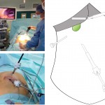
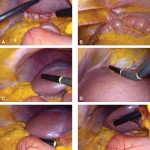
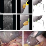
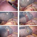
An alternative procedure has come to the fore recently. The ALPPS procedure (Associating Liver Partition and Portal vein Ligation for Staged Liver resection) combines this embolisation procedure with an operation to cut the liver along the line required to remove the diseased portion. But after making the cut, the operation is stopped and the patient woken up. Over the course of the following week the liver being left behind increases in size – quicker and more effectively say proponents of the ALPPS procedure. After a week, the patient is taken back to the operating room and the disease liver portion removed.

So should we start using the procedure to treat cancer which is widely spread in the liver?

The difficulty is knowing whether the new procedure is safe and effective. Early results suggest quite a high mortality associated with the procedure. But of course for patients with untreated cancer in the liver who do not have surgery, the mortality rate is high.

A study has been published which contains some positive data: ALPPS offers a better chance of complete resection in patients with primarily unresectable liver tumors compared with conventional-staged hepatectomies: results of a multicenter analysis.

However, it is still my feeling that the results of the procedure are not good and the traditional portal vein embolisation procedure seems to work well in our patients. Here is our letter with our concerns in response.

We read with interest the multicenter study by Schadde and colleagues in the April issue regarding the novel procedure of Associating Liver Partition and Portal vein Ligation for Staged Liver resection (ALPPS) [1]. Since the initial description 2 years ago [2] ALPPS has gained popularity as a surgical option for treating patients with advanced liver lesions not considered amenable to conventional two-stage or future liver remnant-enhancing procedures propagated by Rene Adam et al. [3] a decade ago. Indeed, the explosion of interest in ALPPS by surgeons and its adoption as a procedure of choice is concerning, given that the procedure appears to come with considerable cost to the patient, as shown in this study. The increased severe morbidity of 27 versus 15 % and the mortality of 15 versus 6 % may not achieve traditional measures of statistical significance in this study, but the effect size is concerning, and the direction of effect is consistent across outcome measures and studies. Is ALPPS in its current form safe enough for the widespread adoption that has occurred given increasingly effective nonsurgical approaches, including ablation, chemotherapy, selective internal radiation therapy [4], and growth factor/receptor inhibition?

As the authors rightly point out, the risk of selection bias is significant given the study design. It is unclear whether the logistic regression analysis adequately adjusts for the imbalance in baseline risk in favor of the ALPPS group: why, for instance, was operative risk (ASA grade) not controlled for in the multivariate analysis?

One of the potential benefits of a two-stage procedure is that it may disclose biologically unfavorable disease. By its very nature, ALPPS does not lend itself to such selection given the short time interval between the first and second stages. The authors appear to reject this argument, citing a similar overall recurrence rate seen in this study. We were puzzled with this position given that the study highlights an interesting observation: in the PVE/PVL group 11 % of patients had systemic progression prior to the second stage. Presumably this group of patients would not have benefitted from ALPPS.

In our practice, patients who may be deemed by others to be ideal candidates for ALPPS are seldom not amenable to either a two-stage liver resection or a single-stage resection with prior volume-enhancing maneuvers. Indeed, it is difficult to understand why an ALPPS approach was used at all in some of the cases presented at recent international conferences. We wonder what proportion and kind of patients with advanced liver lesions would really benefit from the ALPPS approach. The international ALPPS registry will perhaps provide clearer evidence for the role of this challenging approach to liver resection.

1. Schadde E, Ardiles V, Slankamenac K et al (2014) ALPPS offers a better chance of complete resection in patients with primarily unresectable liver tumors compared with conventional-staged hepatectomies: results of a multicenter analysis. World J Surg 38:1510–1519. doi:10.1007/s00268-014-2513-3

2. Schnitzbauer AA, Lang SA, Goessmann H et al (2012) Right portal vein ligation combined with in situ splitting induces rapid left lateral liver lobe hypertrophy enabling 2-staged extended right hepatic resection in small-for-size settings. Ann Surg 255:405–414

3. Adam R, Delvart V, Pascal G et al (2004) Rescue surgery for unresectable colorectal liver metastases downstaged by chemotherapy: a model to predict long-term survival. Ann Surg 240:644–657 discussion 657–658

4. Gulec SA, Pennington K, Wheeler J et al (2013) Yttrium-90 microsphere-selective internal radiation therapy with chemotherapy (chemo-SIRT) for colorectal cancer liver metastases: an in vivo double-arm-controlled phase II trial. Am J Clin Oncol 36:455–460


[gview file=”http://www.datasurg.net/wp-content/uploads/2014/11/Rohatgi-et-al.-2014-ALPPS-Adverse-Outcomes-Demand-Clear-Justification.pdf”]

Hepatitis C virus, tumour and liver transplantation

HCV virus exploding (iStock)

From my HPB highlights this month.

Do patients with hepatocellular carcinoma (HCC) on a background of hepatitis C virus (HCV) have worse outcomes after liver transplantation than non-HCV patients? This relatively straightforward question continues to vex and published studies are contradictory. Molecular features of HCC which are associated with aggressive behaviour are up-regulated in the presence of HCV, providing a biological mechanism to support the hypothesis. The theory is borne out in early single centre studies, but the largest published analysis using the United Network for Organ Sharing database published by Thuluvath in 2009 contradicted these. HCV+ patients were shown to have a lower survival rate than HCV- patients, regardless of their HCC status. This is to be expected. However, HCV had no additional negative impact on survival in patients with HCC

In this edition of HPB, Dumitra and colleagues describe a further single-centre study from Montreal. They conclude that HCC+/HCV+ patients have a significantly worse outcome than those with HCC or HCV alone. So why the contradiction? It may be that length of follow-up is important. This study provides survival curves out to 10 years. A cluster of deaths after 5 years in the HCV+/HCC+ group results in a significantly worse outcome in this group, although the number-at-risk are low. However, loss to follow-up is an unusually low 1.2% and explant pathology is available for almost all patients – detail not often available in studies using administrative databases. In a multivariable analysis controlling for recipient age, gender, MELD score and donor risk index (DRI), the combined effect of HCC+/HCV+ gives a hazard twice that of HCC+/HCV-.

HCV graft infection after liver transplantation is universal and the course of recurrent cirrhosis accelerated. Controlling HCV recurrence with newer antiviral agents will improve long-term survival and this study suggests the possibility of additional benefits in HCC+/HCV+ patients. Other modifiable variables such as donor age and DRI are unlikely to have an impact, given HCC patients rarely have the luxury of a wide choice of donor grafts.

Images in operative ultrasound

Mickey Mouse and the tubes connecting the liver

In liver surgery, it’s often important to know the exact layout of the connections the liver has to the rest of the body. Here are some images which hopefully make it clear. The liver is unusual because it has two blood supplies. The first is an an artery, the hepatic artery, which carries oxygen to the liver. The other is the portal vein which carries blood from the guts to the liver and contains the nutrients from food. The portal vein carries 3 times as much blood as the artery and is not to be messed with – 34% of patients with a portal vein injury do not survive.

The other important tube is the bile duct. This drains bile from the liver to the guts. If it gets blocked – by a gallstone or cancer – the patient becomes jaundiced (the skin going yellow).

We use an ultrasound machine to visualise the vessels and the bile duct. It can be tricky and difficult to interpret. The boss has a good technique for getting orientated – the Mickey Mouse sign. When seen in the transverse plane – imagine sitting at the patient’s feet looking up through the body towards the head – the large portal vein with the artery and bile duct in front looks like Mickey. I use this technique every time.

Intraoperative ultrasound to portal pedicle. Patient consent for publication obtained.
Portal pedicle on ultrasound. CHD, common hepatic duct; LHA, left hepatic artery; RHA, right hepatic artery; PV, portal vein; CBD, common bile duct; PHA, proper hepatic artery; aRHA, accessory right hepatic artery (if present); CHA, common hepatic artery; GDA, gastroduodenal artery; SMV/SV, superior mesenteric vein / splenic vein